This topic covers body organization and how each organ system works. Learn the directional terms, then each system's pathway, main structures, and controlling cells or hormones.
Body organization and directional terms
- Levels, simplest first: chemical (atoms and molecules), cell, tissue (similar cells), organ (two or more tissue types), organ system, organism.
- Anatomical position: standing upright, arms down beside the body, palms facing forward; right and left always mean the patient's own sides.
- Superior/inferior = above/below; anterior/posterior = front/back; superficial/deep = nearer to/farther from the surface.
- Medial means toward the midline and lateral means away from it, so the thumb is lateral to the little finger.
- Proximal means nearer to where a limb joins the trunk and distal means farther: the elbow is proximal to the wrist.
- Planes: sagittal = left/right (midsagittal = equal halves), frontal or coronal = front/back, transverse = upper/lower.
- The dorsal cavity holds only the brain (cranial) and spinal cord (vertebral); the ventral cavity is thoracic plus abdominopelvic, and the kidneys sit behind the peritoneum, not in the dorsal cavity.
- Quadrant lines cross at the navel: the right part of the liver is in the right upper quadrant, the stomach in the left upper, and the appendix in the right lower, where appendicitis pain usually settles after starting near the navel.
Respiratory system
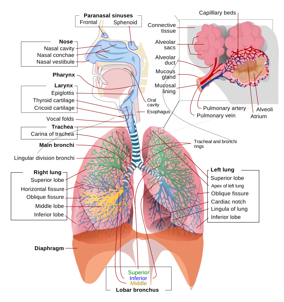
- Air path: nasal cavity → pharynx → larynx → trachea → bronchi → bronchioles → alveoli; gas exchange starts only at the respiratory bronchioles and alveoli.
- C-shaped cartilage rings hold the trachea open; goblet cells make mucus that traps debris, and cilia sweep it toward the throat.
- Each alveolus has a very thin wall wrapped in capillaries, so oxygen and carbon dioxide simply diffuse across.
- Surfactant from type II alveolar cells reduces surface tension, which keeps the alveoli from collapsing at the end of each breath out.
- Inhaling: the diaphragm contracts and moves down, the chest enlarges, lung pressure drops, and air flows in; quiet exhaling is passive.
- The medulla and pons control breathing; rising CO₂ (which lowers blood pH) is the main trigger to breathe harder, and low O₂ becomes a strong trigger only when it falls to about 60 mm Hg or less.
- Nearly all oxygen rides on hemoglobin; carbon dioxide travels about 70% as bicarbonate, about 23% on hemoglobin and about 7% dissolved.
Cardiovascular system and blood
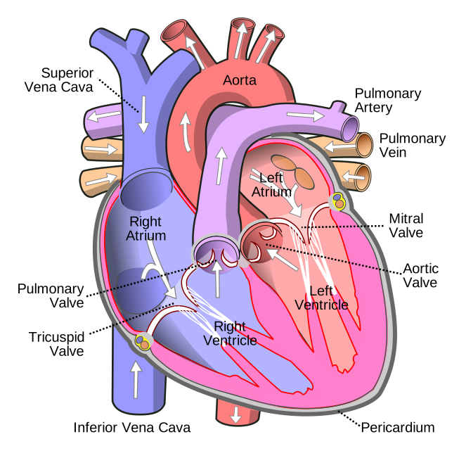
- Blood route: venae cavae → right atrium → tricuspid valve → right ventricle → pulmonary valve → pulmonary arteries → lungs → pulmonary veins → left atrium → mitral (bicuspid) valve → left ventricle → aortic valve → aorta.
- Arteries carry blood away from the heart and veins carry it toward the heart, whatever its oxygen level: pulmonary arteries carry oxygen-poor blood and pulmonary veins oxygen-rich blood.
- Arteries have thicker muscular walls than veins; many leg veins have valves; the veins hold nearly 70% of the blood at any moment; capillaries handle exchange with tissues.
- Conduction: SA node (pacemaker, right atrium) → AV node (brief delay so the atria finish) → bundle of His → bundle branches → Purkinje fibers.
- If the SA node fails, the AV node takes over at about 40–60 beats per minute.
- Type A red cells carry A antigens and the plasma has anti-B antibodies; type B is the reverse; AB has both antigens and neither antibody; O has neither antigen and both antibodies.
- Rh-negative people lack the D antigen and make anti-Rh antibodies only after exposure; O-negative is the universal donor and AB-positive the universal recipient.
Example
Which red cells suit a type B-negative patient: A-negative, AB-negative, B-positive or O-negative?
Solution
B plasma has anti-A antibodies, so rule out A and AB cells. Rh-negative rules out B-positive. O-negative cells carry no A, B or Rh antigen, so they are compatible.
Digestive and urinary systems
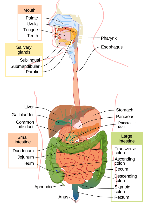
- Food path: mouth → pharynx → esophagus → stomach → small intestine (duodenum, jejunum, ileum) → large intestine (cecum, with the appendix attached) → anus.
- Salivary amylase starts starch digestion in the mouth.
- In the stomach, chief cells release pepsinogen, which hydrochloric acid from parietal cells turns into pepsin to start protein digestion.
- Parietal cells also make intrinsic factor, needed to absorb vitamin B12; the stomach itself absorbs little besides some drugs such as alcohol and aspirin.
- Villi and microvilli give the small intestine a huge surface; nearly all nutrient absorption happens there.
- The liver makes bile, stores glycogen and processes nutrients, drugs and toxins arriving from the intestines; the gallbladder stores and concentrates bile, and bile salts emulsify fat so lipase can digest it.
- Pancreatic juice adds amylase, lipase and trypsinogen (activated to trypsin in the small intestine), released in response to the hormones secretin and CCK.
- The large intestine absorbs water, salts and bacteria-made vitamins such as vitamin K.
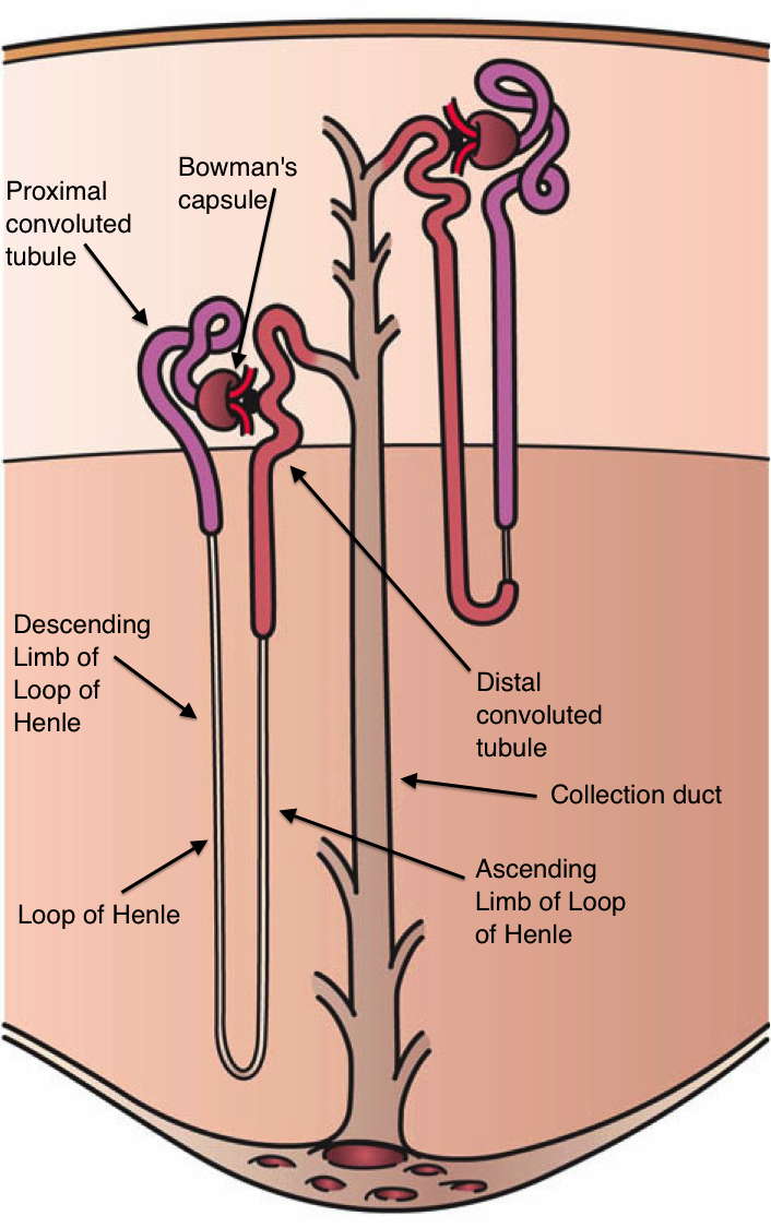
- Nephron (the kidney's functional unit): glomerulus inside Bowman's capsule → proximal tubule → loop of Henle → distal tubule → collecting duct.
- Glomerular blood pressure pushes fluid and small solutes into the capsule; cells and larger proteins stay in the blood.
- The proximal tubule reclaims about two-thirds of the filtered water and sodium and nearly all the glucose.
- In uncontrolled diabetes, filtered glucose exceeds the tubule carriers' maximum, so the extra spills into the urine.
- Urine path: collecting duct → calyces → renal pelvis → ureter → bladder → urethra.
Nervous system and senses
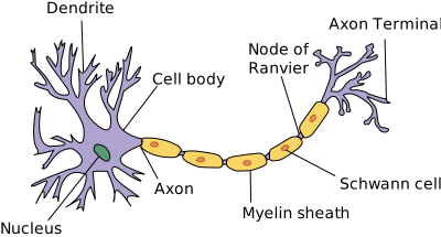
- Dendrites receive signals, and the axon carries impulses away from the cell body.
- Myelin insulates axons and speeds conduction; oligodendrocytes make it in the CNS and Schwann cells make it in the PNS.
- Action potential: from about −70 mV at rest, Na⁺ rushes in through voltage-gated channels (depolarization), then K⁺ flows out (repolarization).
- Cerebral lobes: frontal (movement), parietal (body sensation), temporal (hearing), occipital (vision).
- The thalamus relays every sense except smell, the hypothalamus runs the autonomic system and the anterior pituitary, the cerebellum coordinates movement and balance, and the medulla and pons regulate heart and breathing.
- Somatic nerves control skeletal muscle; autonomic nerves control cardiac muscle, smooth muscle and glands.
- Sympathetic (fight or flight) raises heart and breathing rates, dilates the pupils, reduces saliva (dry mouth), slows digestion and triggers epinephrine release; parasympathetic (rest and digest) mostly does the reverse.
- Reflex arc: receptor → sensory neuron → spinal cord → motor neuron → effector (muscle).
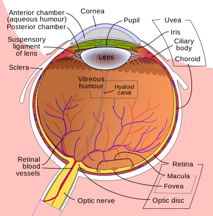
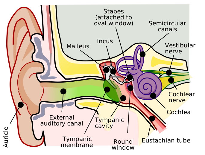
- Cochlear hair cells turn sound into nerve signals, the semicircular canals sense head rotation for balance, and the Eustachian tube links the middle ear to the pharynx.
Muscular, skeletal and integumentary systems
- Skeletal muscle is striated, voluntary and multinucleated; it moves bones, holds posture and makes heat.
- Cardiac muscle is striated, branched and involuntary; intercalated discs with gap junctions let its cells contract together.
- Smooth muscle is unstriated and involuntary and forms the walls of hollow organs such as the intestines and blood vessels.
- Contraction: acetylcholine → Ca²⁺ from the sarcoplasmic reticulum binds troponin → tropomyosin uncovers actin → myosin pulls thin filaments inward; the filaments slide but do not shorten.
- Myosin needs ATP to let go of actin, so cross-bridges stay locked after death (rigor mortis).
- In elbow flexion the biceps is the prime mover and the triceps, which opposes it, is the antagonist.
- Bones give support and protection, hold the body's calcium reserve, form blood cells in red marrow and keep fat in yellow marrow.
- Osteoblasts build bone, osteocytes maintain it, and osteoclasts break it down, releasing calcium.
- Long bones lengthen at the epiphyseal plate until it closes; the periosteum adds width.
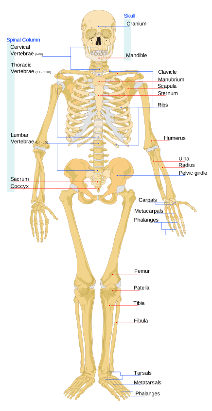
- Axial skeleton (80 bones): skull, vertebrae, ribs, sternum; appendicular skeleton (126): limbs and girdles, including the clavicle and scapula.
- Ball-and-socket joints (shoulder, hip) move on many axes, saddle joints (thumb base) on two, and hinge (elbow, knee) and pivot joints on one.
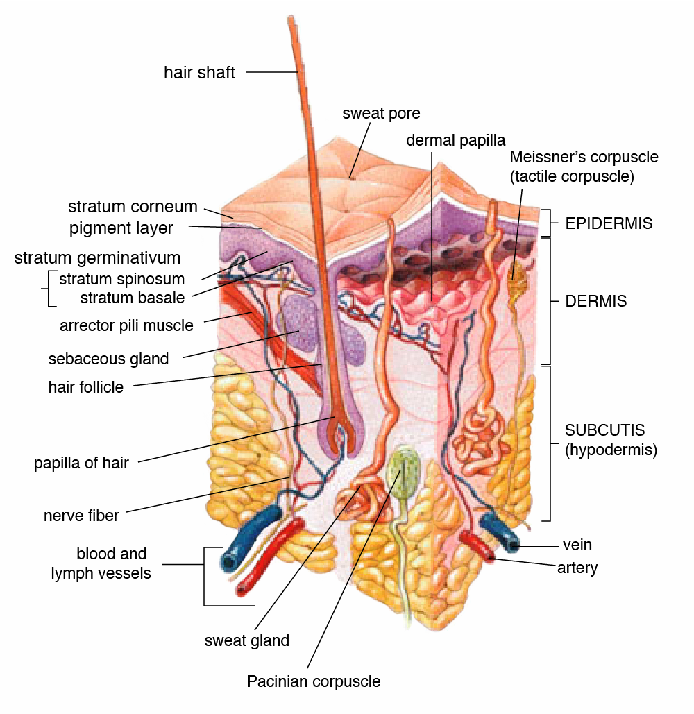
- The epidermis has no blood supply of its own; melanocytes there produce melanin, which shields cell DNA from UV light.
- Sebaceous glands secrete oily sebum; eccrine glands release watery sweat.
- In heat, dermal vessels dilate and sweat cools the skin; in cold, the vessels constrict and goose bumps form.
- Skin also limits water loss and makes vitamin D with UV light.
- Burns: first-degree affects the epidermis only, second-degree reaches the dermis (blisters), and third-degree runs through all skin layers, and the burned skin may be numb, so it can hurt less.
Endocrine system
- The anterior pituitary, controlled by the hypothalamus, makes GH, TSH, ACTH, FSH, LH and prolactin; the posterior pituitary releases ADH and oxytocin made in the hypothalamus.
- Negative feedback keeps levels steady: TRH → TSH → T3 and T4, and high T3 and T4 suppress TRH and TSH, while low levels let them rise.
- Thyroid hormones raise metabolic rate; calcitonin is released when blood calcium rises.
- PTH raises blood calcium by activating osteoclasts, increasing calcium reabsorption in the kidney and activating vitamin D; long-term excess thins the bones.
- Insulin (pancreatic beta cells) lowers blood glucose by moving it into cells and storing it as glycogen; glucagon (alpha cells), cortisol and epinephrine raise it.
- Adrenal cortex: aldosterone (keeps Na⁺, excretes K⁺) and cortisol (long-term stress); adrenal medulla: epinephrine and norepinephrine for sudden stress.
- ADH adds water channels to collecting ducts, giving less, more concentrated urine; atrial natriuretic hormone (ANH, also called atrial natriuretic peptide) increases sodium and water loss.
- Oxytocin drives uterine contractions and milk ejection.
Immune, lymphatic and reproductive systems
- Lymph vessels carry surplus tissue fluid back into the blood, and lymph nodes strain out debris and pathogens along the way.
- B cells finish developing in red bone marrow and T cells in the thymus; lymph nodes, spleen and tonsils host immune responses.
- Neutrophils (50–70% of white cells) are rapid first responders to bacteria; eosinophils fight parasitic worms and allergies; basophils release histamine; monocytes become macrophages.
- Activated B cells become antibody-secreting plasma cells; HIV destroys helper T cells (CD4⁺), which support both antibody and T cell responses.
- Active immunity comes from infection (natural) or a vaccine (artificial); passive immunity comes from a mother's antibodies (natural) or injected antibodies (artificial).
- Sperm path: seminiferous tubules (where sperm form) → epididymis (maturation and storage) → ductus deferens → ejaculatory duct → urethra; seminal vesicles and prostate add fluid.
- FSH grows ovarian follicles, an LH surge triggers ovulation, and the corpus luteum then secretes progesterone.
- Fertilization usually occurs in the ampulla of the uterine tube, and the embryo implants in the endometrium.
- With no implantation, the corpus luteum breaks down, estrogen and progesterone fall, and the endometrium is shed; after implantation, the embryo's hCG keeps the corpus luteum working and is what pregnancy tests detect.
Key terms
- Proximal
- Closer to the point where a limb attaches to the trunk; the opposite of distal.
- Frontal (coronal) plane
- A vertical plane separating the body's front (anterior) part from its back (posterior) part.
- Sagittal plane
- A vertical plane separating the body's right side from its left side.
- Alveoli
- Tiny lung air sacs where oxygen diffuses into the blood and carbon dioxide diffuses out.
- Surfactant
- Phospholipid and protein mix from type II cells that lowers surface tension so air sacs stay open.
- Sinoatrial (SA) node
- Pacemaker tissue in the right atrium wall that sets the normal heart rate.
- Mitral (bicuspid) valve
- The valve between the left atrium and left ventricle.
- Artery
- A vessel carrying blood away from the heart, whether that blood is oxygen-rich or oxygen-poor.
- Intrinsic factor
- Glycoprotein from stomach parietal cells that the small intestine needs to absorb vitamin B12.
- Bile
- Liver secretion whose salts emulsify fat into small droplets for lipase to digest.
- Nephron
- Microscopic working unit of the kidney that filters blood and processes the filtrate into urine.
- Antidiuretic hormone (ADH)
- Posterior pituitary hormone that opens water channels in collecting ducts, producing less, more concentrated urine.
- Aldosterone
- Adrenal cortex hormone that makes the kidneys keep sodium and excrete potassium.
- Negative feedback
- A control loop in which a rising output switches off its own stimulus, keeping levels steady.
- Osteoclast
- Large, multinucleated bone cell that breaks down bone matrix and releases calcium into the blood.
- Epiphyseal plate
- Cartilage growth zone near each end of a long bone where it lengthens until growth ends.
- Axon
- Long neuron extension that conducts impulses away from the cell body.
- Antagonist
- A muscle that works against the prime mover, producing the opposite movement.
- Plasma cell
- Cell that develops from an activated B lymphocyte and releases antibodies in large amounts.
- Human chorionic gonadotropin (hCG)
- Embryo hormone that keeps the corpus luteum active; home pregnancy tests detect it.
Common mistakes
- Calling every artery oxygen-rich. Arteries are named by direction (away from the heart), so the pulmonary arteries carry oxygen-poor blood and the pulmonary veins carry oxygen-rich blood.
- Reversing the diaphragm. It contracts and flattens to pull air in; in quiet breathing it relaxes to let air out.
- Saying bile digests fat. Bile only emulsifies fat into droplets; the enzyme lipase does the chemical splitting.
- Judging direction from your own point of view. Right, left, medial and lateral are judged on the patient in anatomical position, so the thumb is lateral and the appendix lies in the right lower quadrant.
- Getting feedback backward. Too much thyroid hormone lowers TSH, and long-term excess PTH pulls calcium out of bone, raising blood calcium and thinning bone.
- Mixing up kidney controls. ADH directly adds water channels, aldosterone retains sodium, and glucose in the urine in diabetes comes from saturated tubule carriers, not leaky glomeruli.
Checked against: MedlinePlus, Appendicitis; Wikipedia, Biological organisation; Wikipedia, Tissue (biology); Wikipedia, Anatomical terms of location; Wikipedia, Standard anatomical position; Wikipedia, Anatomical plane; Wikipedia, Body cavity; Wikipedia, Retroperitoneal space; Wikipedia, Quadrants and regions of abdomen; Wikipedia, Respiratory tract; Wikipedia, Trachea; Wikipedia, Pulmonary alveolus; Wikipedia, Pulmonary surfactant; Wikipedia, Thoracic diaphragm; Wikipedia, Breathing; Wikipedia, Control of ventilation; Wikipedia, Carotid body; Wikipedia, Blood; Wikipedia, Heart; Wikipedia, Electrical conduction system of the heart; Wikipedia, Artery; Wikipedia, Pulmonary artery; Wikipedia, Vein; Wikipedia, Capillary; Wikipedia, ABO blood group system; Wikipedia, Rh blood group system; Wikipedia, Blood type; Wikipedia, Human digestive system; Wikipedia, Gastric chief cell; Wikipedia, Parietal cell; Wikipedia, Stomach; Wikipedia, Liver; Wikipedia, Bile; Wikipedia, Pancreas; Wikipedia, Large intestine; Wikipedia, Nephron; Wikipedia, Proximal tubule; Wikipedia, Glycosuria; Wikipedia, Urinary system; Wikipedia, Neuron; Wikipedia, Action potential; Wikipedia, Lobes of the brain; Wikipedia, Thalamus; Wikipedia, Hypothalamus; Wikipedia, Cerebellum; Wikipedia, Medulla oblongata; Wikipedia, Autonomic nervous system; Wikipedia, Fight-or-flight response; Wikipedia, Reflex arc; Wikipedia, Inner ear; Wikipedia, Eustachian tube; Wikipedia, Skeletal muscle; Wikipedia, Cardiac muscle; Wikipedia, Muscle tissue; Wikipedia, Smooth muscle; Wikipedia, Muscle contraction; Wikipedia, Anatomical terms of muscle; Wikipedia, Bone; Wikipedia, Osteocyte; Wikipedia, Periosteum; Wikipedia, Epiphyseal plate; Wikipedia, Axial skeleton; Wikipedia, Appendicular skeleton; Wikipedia, Synovial joint; Wikipedia, Saddle joint; Wikipedia, Hinge joint; Wikipedia, Pivot joint; Wikipedia, Epidermis; Wikipedia, Melanin; Wikipedia, Human skin; Wikipedia, Eccrine sweat gland; Wikipedia, Goose bumps; Wikipedia, Burn; MedlinePlus, Burns; Wikipedia, Pituitary gland; Wikipedia, Hypothalamic–pituitary–thyroid axis; Wikipedia, Thyroid hormones; Wikipedia, Calcitonin; Wikipedia, Parathyroid hormone; MedlinePlus, Hyperparathyroidism; Wikipedia, Blood sugar regulation; Wikipedia, Adrenal gland; Wikipedia, Cortisol; Wikipedia, Vasopressin; Wikipedia, Atrial natriuretic peptide; Wikipedia, Oxytocin; Wikipedia, Lymphatic system; Wikipedia, White blood cell; Wikipedia, Neutrophil; Wikipedia, Plasma cell; Wikipedia, T helper cell; Wikipedia, Immunity (medicine); Wikipedia, Male reproductive system; Wikipedia, Epididymis; Wikipedia, Ejaculatory duct; Wikipedia, Menstrual cycle; Wikipedia, Fertilisation; Wikipedia, Human chorionic gonadotropin