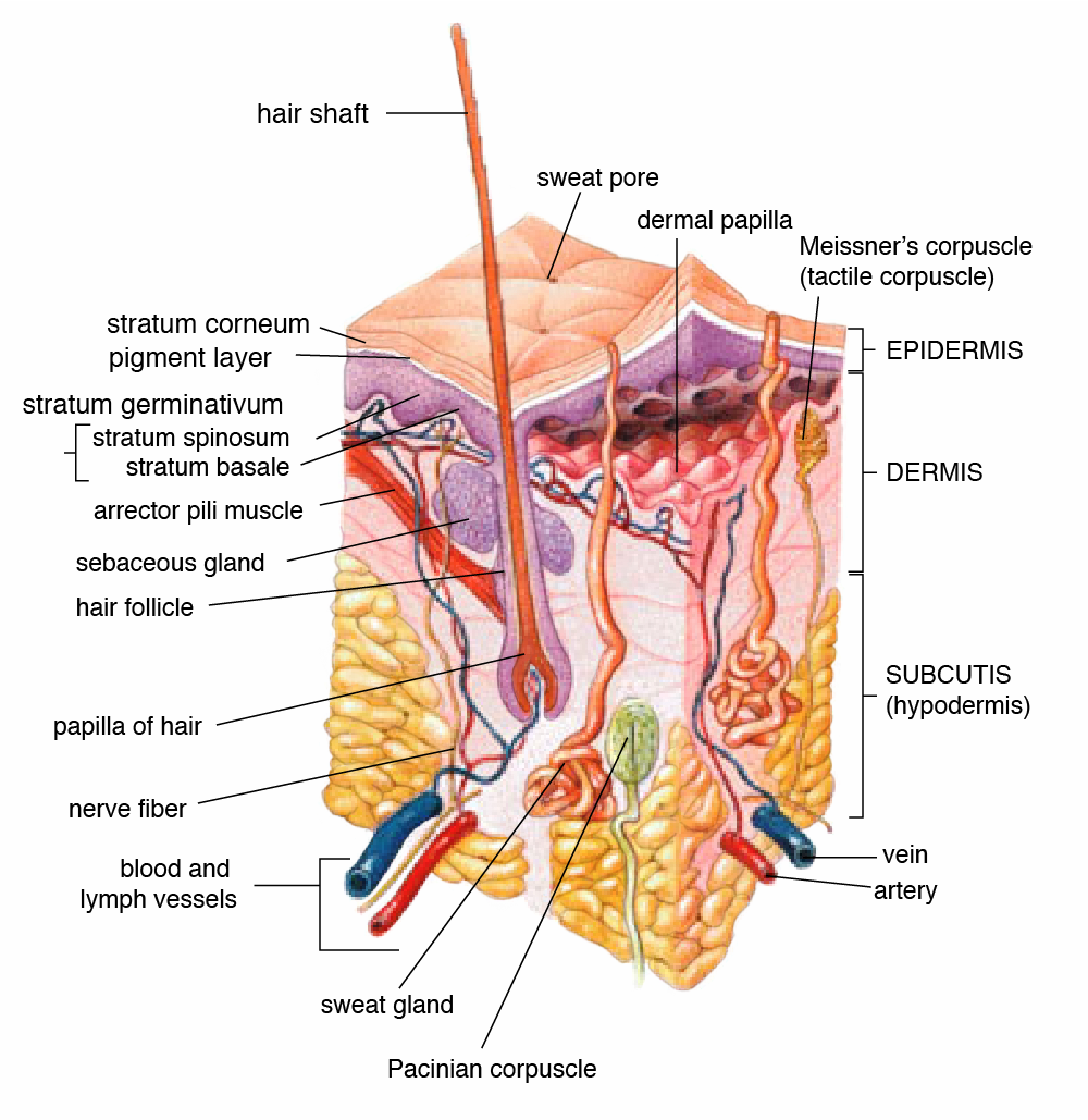
This topic covers the shared vocabulary of anatomy (position, directions, planes and cavities), the levels of organization, homeostasis, the four tissue types and the layers of the skin. Learn directional terms as opposite pairs, and link each tissue to where it is found and why.
Anatomical position and directional terms
Directional terms always refer to the body in anatomical position, no matter how the person is actually lying or standing.
- Anatomical position: upright and facing forward, feet parallel, arms hanging by the sides with the palms facing forward.
- Supine is lying on the back, face up; prone is lying on the front, face down; neither one is anatomical position.
- Superior (cranial) describes a part higher up, toward the head; inferior (caudal) describes a part lower down, toward the feet.
- Anterior (ventral) refers to the front of the body; posterior (dorsal) refers to the back.
- Medial points toward the body's midline; lateral points away from it, toward the side.
- Proximal means closer to the trunk or to where a limb attaches, and distal means farther away, so the elbow is proximal to the wrist.
- Superficial means nearer the body surface and deep means farther in, so the skin is superficial to the muscles.
Body planes and cavities
- A sagittal plane divides the body into right and left parts; the midsagittal (median) plane passes through the midline and gives equal halves.
- A frontal (coronal) plane divides the body into anterior (front) and posterior (back) parts.
- A transverse (horizontal) plane divides the body into superior (upper) and inferior (lower) parts, giving a cross section.
- The dorsal cavity is made up of the cranial cavity, which holds the brain, and the vertebral (spinal) cavity, which holds the spinal cord.
- The diaphragm splits the ventral cavity into the thoracic cavity above it and the abdominopelvic cavity below it.
- Within the thoracic cavity, each lung sits in a pleural cavity of its own, and the heart sits in the pericardial cavity inside the mediastinum, the central space between the lungs.
- Digestive organs occupy the abdominal cavity; the reproductive organs and the bladder occupy the pelvic cavity.
- Serous membranes (pleura, pericardium and peritoneum) line these cavities and wrap their organs, and the serous fluid between the layers cuts down friction.
Levels of organization
- Chemical level: the atoms and molecules from which everything in the body is built.
- Cellular level: the cell, the smallest part of the body that can carry out life's functions on its own.
- Tissue level: similar cells grouped together to carry out a shared task.
- Organ level: a structure in which two or more kinds of tissue work as one unit.
- Organ system level: a set of organs that together handle a major body function.
- Organism level: the complete living person, formed by all the organ systems working together.
Example
Put these in order from simplest to most complex: tissue, organism, chemical, organ system, cellular, organ.
Solution
Chemical → cellular → tissue → organ → organ system → organism. Molecules make up cells, similar cells group into tissues, different tissues combine into an organ, organs team up as a system, and the systems together form the organism.
Homeostasis and feedback loops
Homeostasis means holding the body's internal conditions within a normal range around a set point. For body temperature, the widely used set point is about 37 °C (98.6 °F).
- Sensor (receptor): picks up the change and sends the information on.
- Control center: checks the reading against the normal range and issues commands.
- Effector: the muscle or gland that produces the correcting response.
- Negative feedback undoes a change, as with sweating when overheated, shivering when chilled, and insulin release after blood glucose rises.
- Positive feedback pushes a change further until some end point; childbirth and blood clotting are normal examples.
- During labor, pressure of the baby on the cervix sends signals to the brain, the pituitary releases oxytocin, and contractions strengthen until the birth stops the loop.
Example
Body temperature falls and the person starts to shiver. Identify the stimulus, sensor, control center and effector.
Solution
Stimulus: the fall in temperature. Sensor: temperature receptors in the skin and brain. Control center: the brain's temperature-regulating center. Effector: the skeletal muscles, which shiver and produce heat. The response undoes the fall, so this is negative feedback.
The four tissue types
- Epithelial tissue covers body surfaces, lines cavities and passages, and forms glands; it has no blood vessels of its own.
- Connective tissue holds other tissues together and supports and protects them; its three groups are connective tissue proper (loose, adipose, and dense types such as tendons and ligaments), supportive (cartilage and bone) and fluid (blood and lymph).
- Muscle tissue contracts to create movement and has three types: skeletal, cardiac and smooth.
- Nervous tissue, made of neurons and glia, detects stimuli and conducts electrochemical signals.
- Epithelium is named for its number of layers (simple = one, stratified = more than one) and for its cell shape (squamous = flat, cuboidal = cube-like, columnar = tall).
- Simple squamous epithelium is thin enough for gases to cross it quickly, so it lines the alveoli and the capillaries.
- Stratified squamous epithelium stands up to wear: the keratinized type forms the outer skin, and the nonkeratinized type lines the mouth and esophagus.
- Simple columnar epithelium lines the stomach and intestines, and ciliated pseudostratified columnar epithelium lines the trachea and bronchi.
- Transitional epithelium, which changes shape as it stretches, lines the ureters and urinary bladder.
The skin
- The epidermis, the outer layer, is keratinized stratified squamous epithelium.
- The dermis beneath it is connective tissue holding blood and lymph vessels, nerves, hair follicles and sweat glands.
- The hypodermis is loose connective and adipose tissue that anchors the skin to deeper tissue, stores fat and insulates.
- The epidermis has no blood vessels, so a scrape bleeds only once it reaches the dermis.
- The epidermis has five layers. Starting at the bottom: stratum basale, then spinosum, then granulosum, then lucidum (found only in the thick skin of palms and soles), and finally corneum on the surface.
- Stem cells in the stratum basale divide to form new keratinocytes, which are pushed upward, fill with keratin and die.
- The stratum corneum consists of many layers of dead, keratin-filled cells that flake off, keep out microbes and hold in water.
- Keratinocytes make keratin, melanocytes make melanin, Merkel cells sense light touch, and Langerhans cells (once classed as dendritic cells) are immune cells that take up microbes.
- Melanocytes pass melanin to keratinocytes, where it absorbs UV radiation and shields the DNA of living epidermal cells.
Key terms
- Anatomical position
- Upright, facing forward, feet parallel, arms at the sides with palms forward; the reference for all directional terms.
- Supine
- Lying face up.
- Prone
- Lying face down.
- Proximal
- Nearer the trunk or the point where a limb attaches; its opposite is distal.
- Superficial
- Closer to the body surface; its opposite is deep.
- Sagittal plane
- A vertical cut that separates the right and left sides of the body.
- Frontal (coronal) plane
- A vertical cut that separates the front of the body from the back.
- Transverse plane
- A horizontal cut that separates upper from lower parts and gives a cross section.
- Mediastinum
- Central region of the thoracic cavity, between the two pleural cavities, that holds the heart.
- Homeostasis
- Keeping internal conditions within a normal range around a set point.
- Negative feedback
- A response that reverses a change and returns a value toward its set point.
- Positive feedback
- A response that strengthens the original change until an end point, as in childbirth.
- Effector
- The muscle or gland in a feedback loop that produces the corrective response.
- Simple squamous epithelium
- One layer of thin, flat cells that allows fast diffusion, as in alveoli and capillaries.
- Connective tissue
- Binds, supports and protects other structures; examples are fat, cartilage, bone, blood and lymph.
- Avascular
- Having no blood vessels; true of the epidermis and of cartilage.
- Stratum basale
- Deepest epidermal layer, where stem cells divide to produce new keratinocytes.
- Melanocyte
- Epidermal cell that makes melanin, the pigment that absorbs UV radiation.
Common mistakes
- Anatomical position has the palms facing forward, not toward the thighs, and the person stands; lying face up (supine) or face down (prone) is not anatomical position.
- Proximal and distal compare points along a limb; to describe nearness to the body surface, use superficial and deep.
- A frontal (coronal) plane separates front from back, while a sagittal plane separates left from right and a transverse plane separates upper from lower.
- The lungs are not in the mediastinum: each lung has its own pleural cavity, and the heart sits in the mediastinum between them.
- Sweating, shivering and insulin release all reverse a change, so they are negative feedback; in a loop the sensor detects, the control center decides, and the effector (such as shivering muscle) acts, while the change itself is the stimulus.
- Blood, bone and adipose tissue are connective tissues, but cardiac muscle is muscle tissue; and melanin comes from melanocytes, not keratinocytes, while new cells come from the stratum basale, not the stratum corneum.
Checked against: Wikipedia, Standard anatomical position; Wikipedia, Anatomical terms of location; Wikipedia, Supine position; Wikipedia, Transverse plane; Wikipedia, Body cavity; Wikipedia, Pleural cavity; Wikipedia, Mediastinum; Wikipedia, Serous membrane; Wikipedia, Biological organisation; Wikipedia, Cell (biology); Wikipedia, Tissue (biology); Wikipedia, Organ (biology); Wikipedia, Homeostasis; Wikipedia, Human body temperature; Wikipedia, Positive feedback; Wikipedia, Epithelium; Wikipedia, Capillary; Wikipedia, Trachea; Wikipedia, Connective tissue; Wikipedia, Cartilage; Wikipedia, Muscle tissue; Wikipedia, Nervous tissue; Wikipedia, Epidermis; Wikipedia, Dermis; Wikipedia, Subcutaneous tissue; Wikipedia, Keratinocyte; Wikipedia, Melanocyte; Wikipedia, Merkel cell; Wikipedia, Langerhans cell