This topic covers blood and its cells, the path of blood through the heart, the heart's electrical system and sounds, blood vessels, the lymphatic and immune systems, and how the lungs move air and exchange gases. Be able to trace blood and air step by step, and know which cell or structure does each job.
Blood
- Plasma, mostly water, is about 55% of blood; red blood cells (the hematocrit) are about 45%; white cells and platelets are under 1%.
- Erythrocytes (red blood cells) are flexible biconcave discs; mature ones lack a nucleus and are packed with oxygen-carrying hemoglobin.
- Each hemoglobin has four globin chains and four iron-containing heme groups, so it can carry four O₂ molecules.
- Red blood cells live about 120 days, and the kidneys release erythropoietin (EPO) to boost their production when oxygen is low.
- Neutrophils are the most common white blood cell (about 40–70%); they are first responders that engulf and digest bacteria.
- Lymphocytes (about 20–40%) are B cells, T cells and natural killer cells; B and T cells carry out specific immunity.
- Monocytes leave the blood and become macrophages; eosinophils fight parasites and allergic reactions; basophils release histamine.
- Platelets are pieces of megakaryocyte cytoplasm that clump into a plug where a vessel is torn, the key step in hemostasis (stopping bleeding).
Path of blood through the heart
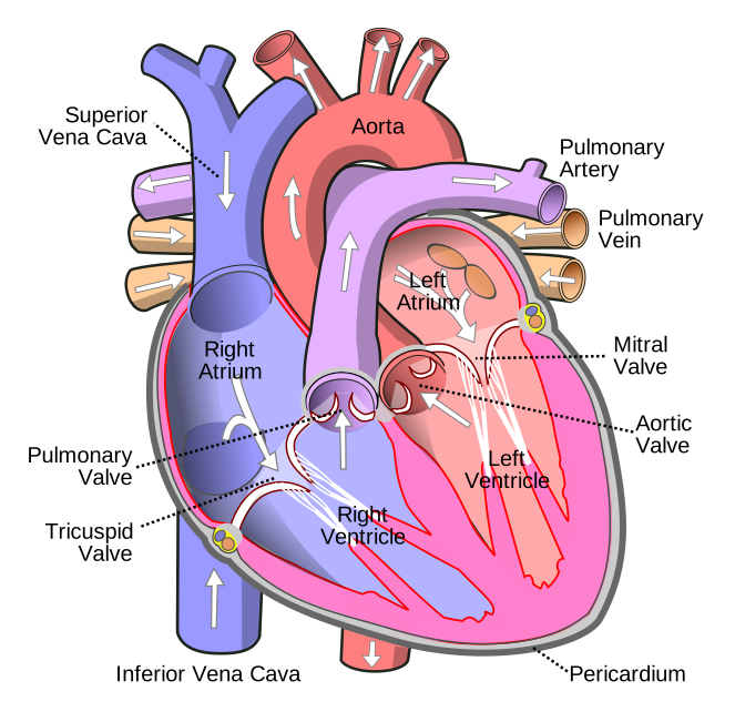
The right side pumps oxygen-poor blood to the lungs; the left side pumps oxygen-rich blood to the body. The left ventricle has the thicker wall because it must push blood through the whole systemic circuit.
- Superior and inferior venae cavae → right atrium → tricuspid valve → right ventricle → pulmonary valve → pulmonary trunk and arteries → lungs.
- Lungs → pulmonary veins → left atrium → mitral (bicuspid) valve → left ventricle → aortic valve → aorta → body.
- Pulmonary arteries carry oxygen-poor blood to the lungs, and pulmonary veins are the only veins after birth that carry oxygen-rich blood.
- The coronary arteries branch from the aorta and supply the heart muscle itself.
Electrical system and heart sounds
- Conduction pathway: SA node → atria → AV node → AV bundle (bundle of His) → right and left bundle branches → Purkinje fibers.
- The SA node is the pacemaker because its cells depolarize on their own faster than any other part of the heart.
- The AV node holds the impulse back for about 0.09 second (roughly a tenth of a second) so the atria finish emptying into the ventricles before the ventricles contract.
- On an ECG, the P wave is atrial depolarization, the QRS complex is ventricular depolarization and the T wave is ventricular repolarization.
- Systole is contraction and diastole is relaxation and filling.
- S1 ("lub") is the atrioventricular valves (tricuspid and mitral) closing at the start of ventricular systole.
- S2 ("dub") is the semilunar valves (pulmonary and aortic) closing as the ventricles relax.
Blood vessels
- Arteries carry blood away from the heart; layers of smooth muscle and elastic tissue in their walls let them withstand high pressure.
- Arterioles act as adjustable nozzles: tightening or relaxing their smooth muscle sets blood pressure and flow into the capillaries.
- A capillary wall is a single thin layer of endothelial cells, so gases, nutrients and wastes can pass between blood and tissues.
- Veins return blood to the heart under low pressure and hold most of the blood (nearly 70%) at any moment.
- Many veins, especially in the limbs, have valves that keep blood moving toward the heart and stop backflow.
Lymphatic and immune systems
- Lymphatic vessels return leaked tissue fluid (lymph) to the blood, draining into the subclavian veins through the thoracic duct and right lymphatic duct.
- Lacteals in the small intestine absorb dietary fats into the lymph.
- B cells mature in the red bone marrow; T cells form in the marrow but mature in the thymus.
- Lymph nodes filter lymph, and the spleen filters blood and removes old red blood cells.
- Innate defenses slow an infection while the adaptive response builds; adaptive immunity uses B and T cells, is highly specific, and has memory.
- An activated B cell multiplies into plasma cells, which secrete antibodies, and memory B cells, which allow a faster, stronger response next time.
- Helper T cells release cytokines that activate B cells and other immune cells; cytotoxic T cells kill infected cells.
- IgM is usually the first antibody made, IgG crosses the placenta, IgA protects secretions such as tears and breast milk, and IgE is linked to allergy.
Airways and breathing
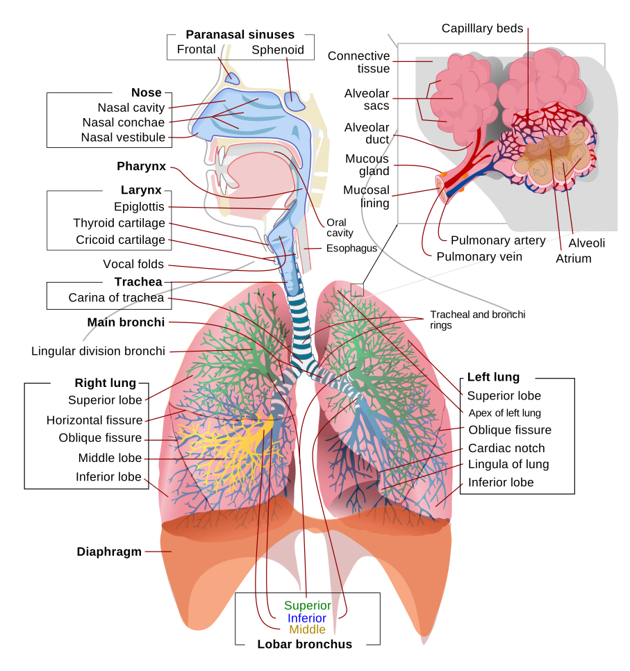
- The conducting zone (nose, pharynx, larynx, trachea, bronchi, bronchioles down to the terminal bronchioles) cleans, warms and moistens air but exchanges no gas.
- Oxygen and carbon dioxide are swapped with the blood only in the respiratory zone (respiratory bronchioles, alveolar ducts and alveoli).
- During swallowing the epiglottis folds over the entrance to the trachea, so food goes down the esophagus instead.
- Type I alveolar cells cover almost all of the alveolar surface, and type II cells release surfactant, without which the alveoli would collapse.
- When the diaphragm contracts and the external intercostals lift the ribs, chest volume rises, lung pressure falls below air pressure, and air flows in (Boyle's law).
- Quiet exhalation is passive elastic recoil; forced exhalation uses the internal intercostals and abdominal muscles.
Gas exchange and gas transport
- The alveolar wall and the capillary wall are fused into one thin respiratory membrane, which gases cross by simple diffusion, using no energy.
- Each gas diffuses down its own partial pressure gradient, from higher to lower, regardless of the other gases.
- About 98.5% of oxygen travels bound to hemoglobin; only about 1.5% dissolves in plasma.
- About 70% of carbon dioxide travels as bicarbonate, about 23% bound to the globin part of hemoglobin, and about 7% dissolved in plasma.
- In red blood cells, carbonic anhydrase speeds CO₂ + H₂O → carbonic acid → H⁺ + bicarbonate, and bicarbonate leaves the cell in exchange for chloride (the chloride shift).
- More CO₂ and acid lower hemoglobin's grip on oxygen (the Bohr effect), so active tissues receive more oxygen.
Example
Alveolar air has a PO₂ of about 100 mm Hg and a PCO₂ of about 40 mm Hg. Blood arriving in the pulmonary capillaries has a PO₂ of about 45 mm Hg and a PCO₂ of about 45 mm Hg. Which way does each gas move?
Solution
Oxygen: 100 mm Hg in the alveoli is higher than 45 mm Hg in the blood, so O₂ diffuses into the blood (gradient 100 − 45 = 55 mm Hg). Carbon dioxide: 45 mm Hg in the blood is higher than 40 mm Hg in the alveoli, so CO₂ diffuses into the alveoli to be exhaled (gradient 45 − 40 = 5 mm Hg).
Key terms
- Platelets
- Megakaryocyte fragments that clump to seal a damaged vessel and stop bleeding.
- Neutrophil
- Most abundant white blood cell; a fast responder that engulfs bacteria.
- Hemoglobin
- Iron-containing protein in red blood cells that binds up to four oxygen molecules.
- Pulmonary veins
- Vessels carrying oxygen-rich blood from the lungs to the left atrium.
- Tricuspid valve
- Right atrioventricular flaps that stop backflow from the right ventricle into the right atrium.
- Mitral (bicuspid) valve
- Left atrioventricular flaps that stop backflow from the left ventricle into the left atrium.
- Sinoatrial (SA) node
- Pacemaker of the heart, in the right atrial wall, that starts each beat.
- Atrioventricular (AV) node
- Conduction tissue that delays the impulse so the atria finish contracting first.
- Systole
- The contraction phase of a heart chamber.
- Diastole
- The relaxation phase, when a heart chamber fills with blood.
- Vein
- Low-pressure vessel returning blood toward the heart, often with one-way valves.
- Capillary
- Microscopic vessel with a one-cell-layer wall, where blood and tissues trade gases, nutrients and wastes.
- Thymus
- Organ in the chest where T lymphocytes mature.
- Plasma cell
- Antibody-secreting descendant of an activated B lymphocyte.
- Memory B cell
- Long-lived lymphocyte that allows a faster, stronger response on later exposure to the same antigen.
- Alveoli
- Tiny sacs at the airway tips where oxygen enters and carbon dioxide leaves the blood.
- Surfactant
- Secretion of type II alveolar cells that lowers surface tension and keeps alveoli open.
- Partial pressure
- How much one gas in a mixture contributes to the total, in mm Hg; each gas diffuses from high to low.
- Bicarbonate ion
- HCO₃⁻; the form in which about 70% of carbon dioxide travels in blood.
Common mistakes
- Platelets, not red or white blood cells, drive hemostasis; and neutrophils (40–70%), not lymphocytes (20–40%), are the most common white blood cells.
- Pulmonary arteries carry oxygen-poor blood and pulmonary veins carry oxygen-rich blood; name a vessel by direction (artery = away from the heart), not by oxygen content.
- The SA node sets the pace; the AV node only delays the impulse so the atria can finish contracting before the ventricles do.
- S1 is the tricuspid and mitral (AV) valves closing; S2 is the aortic and pulmonary (semilunar) valves closing. Do not pair one valve of each type.
- T cells are made in the bone marrow but mature in the thymus; plasma cells, not helper T cells or memory B cells, secrete antibodies.
- Gas exchange is passive diffusion down each gas's own partial pressure gradient; most CO₂ travels as bicarbonate, and the CO₂ that binds hemoglobin attaches to globin, not to the iron in heme.
Checked against: Wikipedia, Blood; Wikipedia, Red blood cell; Wikipedia, Hemoglobin; Wikipedia, Erythropoietin; Wikipedia, White blood cell; Wikipedia, Neutrophil; Wikipedia, Lymphocyte; MedlinePlus Medical Encyclopedia, Blood differential test; Wikipedia, Platelet; Wikipedia, Heart; Wikipedia, Pulmonary artery; Wikipedia, Pulmonary vein; Wikipedia, Sinoatrial node; Wikipedia, Atrioventricular node; Wikipedia, Electrocardiography; Wikipedia, Heart sounds; Wikipedia, Artery; Wikipedia, Capillary; Wikipedia, Vein; Wikipedia, Lymphatic system; Wikipedia, Adaptive immune system; Wikipedia, T helper cell; Wikipedia, Antibody; Wikipedia, Respiratory tract; Wikipedia, Epiglottis; Wikipedia, Pulmonary alveolus; Wikipedia, Breathing; Wikipedia, Intercostal muscle; Wikipedia, Inhalation; Wikipedia, Gas exchange; Wikipedia, Pulmonary gas pressures; Wikipedia, Partial pressure; Wikipedia, Carbaminohemoglobin; Wikipedia, Chloride shift