This topic covers where each nutrient is digested and absorbed, what the liver, gallbladder and pancreas add, how the nephron turns blood plasma into urine and which hormones adjust it, and the main structures and hormones of the male and female reproductive systems. Follow food, filtrate and gametes along their paths and know what happens at each stop.
Mouth to stomach
- In the mouth, chewing breaks food apart and salivary amylase begins digesting starch.
- During swallowing, the epiglottis folds over the airway so food enters the esophagus.
- The esophagus only moves food to the stomach by peristalsis (waves of muscle contraction); it neither digests nor absorbs.
- Parietal cells of the gastric glands make hydrochloric acid as well as intrinsic factor, which is needed to absorb vitamin B12.
- Chief cells secrete pepsinogen, which stomach acid converts to pepsin, so protein digestion begins in the stomach.
- G cells release the hormone gastrin, which increases acid secretion; mucous neck cells secrete mucus.
- Stomach contents are very acidic (pH about 1 to 3), and little is absorbed there apart from some alcohol and drugs such as aspirin.
- The stomach releases chyme to the duodenum in small amounts through the pyloric sphincter.
Small and large intestines
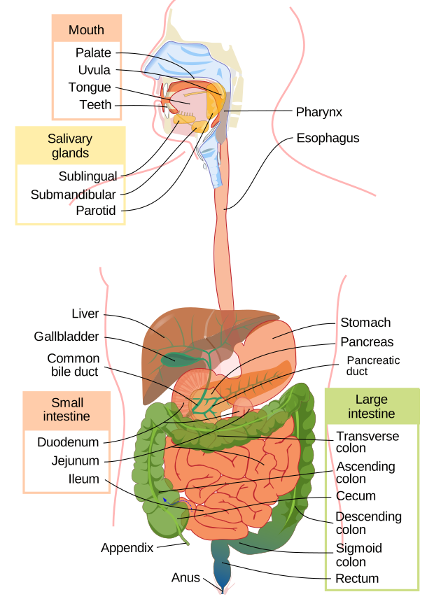
- The small intestine has three parts: duodenum, jejunum and ileum.
- Most chemical digestion and nearly all nutrient absorption happen in the small intestine.
- Circular folds, villi and microvilli (the brush border) give the small intestine an enormous absorptive surface.
- Each villus contains blood capillaries and a lacteal, a lymphatic capillary.
- The large intestine (cecum, colon, rectum) absorbs most of the remaining water and salts and forms feces.
- Bacteria in the large intestine help break down residue and make some vitamins, including vitamin K.
Liver, gallbladder and pancreas
- The liver makes bile; the gallbladder only stores and concentrates it and releases it into the duodenum after a fatty meal.
- Bile salts emulsify fat: they split big fat globules into small droplets so that lipase can get at them.
- Pancreatic juice contains bicarbonate, which neutralizes acidic chyme, and enzymes: amylase (starch), lipase (fat), nucleases (nucleic acids) and inactive protein-digesting enzymes such as trypsinogen.
- Blood leaving the intestines, loaded with absorbed nutrients, goes first to the liver through the hepatic portal vein.
Absorbing the nutrients
- Carbohydrates are absorbed as monosaccharides such as glucose, proteins as amino acids, and fats as fatty acids and monoglycerides.
- Monosaccharides and amino acids enter the blood capillaries of the villi and travel to the liver in the hepatic portal vein.
- Long-chain fatty acids and monoglycerides are rebuilt into triglycerides inside intestinal cells and packed into chylomicrons.
- Chylomicrons pass into the lacteals rather than the blood capillaries, and reach the blood later through the thoracic duct.
- Vitamins A, D, E and K dissolve in fat, so they are absorbed together with dietary fat.
The nephron and urine formation
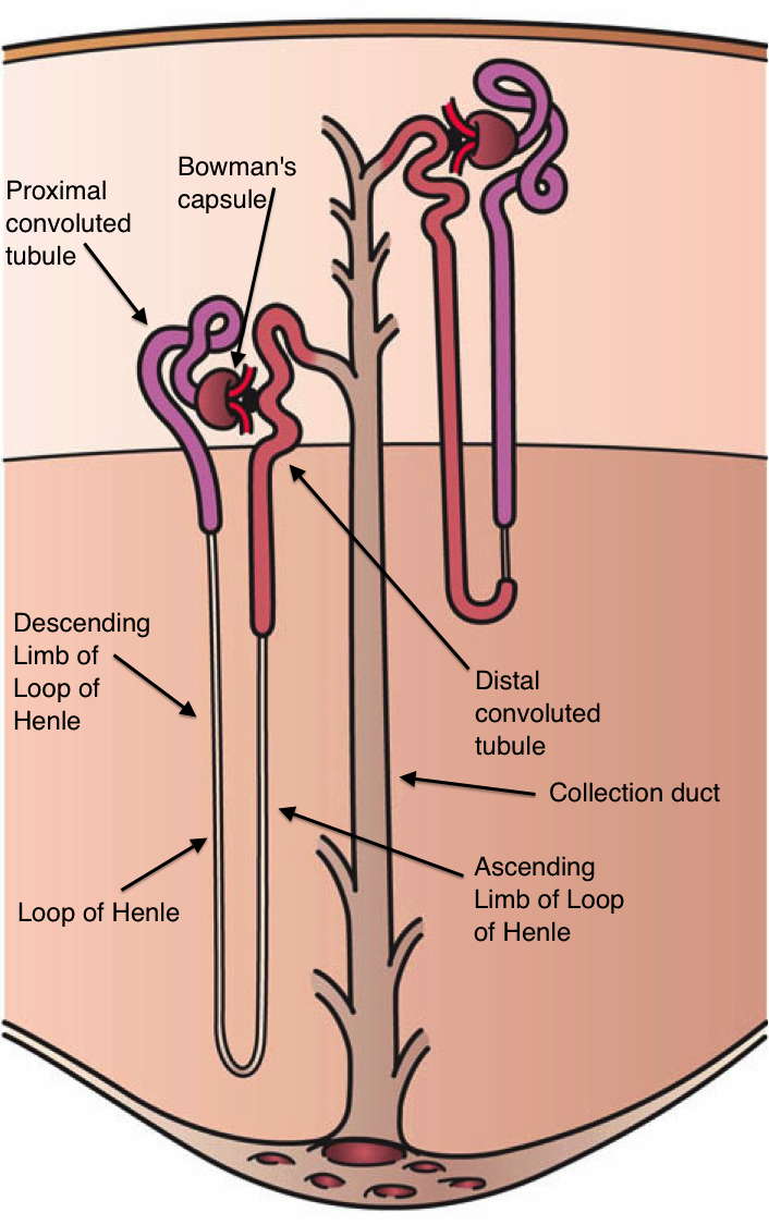
Filtrate flows through the nephron in this order: glomerulus inside the glomerular (Bowman's) capsule → proximal convoluted tubule → nephron loop (loop of Henle) → distal convoluted tubule → collecting duct. Urine then drains through the renal pelvis, ureter, bladder and urethra.
- Filtration happens at the glomerulus: blood pressure pushes water and small solutes into the capsule, while blood cells and large proteins such as albumin stay in the blood.
- The proximal convoluted tubule reabsorbs the largest share, about two-thirds of the filtered water, sodium and potassium.
- The proximal tubule also reabsorbs nearly 100% of filtered glucose and amino acids, so normal urine has no glucose.
- Glucose spills into urine only when blood levels exceed what the transporters can carry, as in uncontrolled diabetes mellitus.
- About half of filtered urea is reabsorbed, while creatinine is not reabsorbed at all.
- Hydrogen ions, potassium and some drugs are secreted from the blood into the tubule to be excreted.
- The nephron loop builds a salt gradient in the kidney's medulla that lets the collecting duct concentrate urine.
- About 99% of the filtrate is reabsorbed, so a person makes only about 1 to 2 liters of urine a day.
Hormones that adjust the kidneys
- ADH (from the posterior pituitary) inserts aquaporin water channels into collecting duct cells, so more water is reabsorbed and urine becomes concentrated.
- Aldosterone (from the adrenal cortex) increases sodium reabsorption and potassium excretion; water follows the sodium, raising blood volume.
- When blood volume falls, the kidneys release renin, which leads to angiotensin II; angiotensin II constricts vessels and triggers aldosterone release.
- Atrial natriuretic peptide (ANP) from the stretched atria does the opposite, so more sodium and water leave in the urine.
Male reproductive system
- Sperm are produced in the coiled seminiferous tubules inside each testis.
- Sertoli (sustentacular) cells inside the tubules support developing sperm and respond to FSH.
- Leydig (interstitial) cells between the tubules make testosterone when stimulated by LH.
- Sperm path: seminiferous tubules → epididymis (sperm mature and gain the potential to swim) → ductus (vas) deferens → ejaculatory duct → urethra.
- The seminal vesicles, prostate and bulbourethral glands add the fluids of semen; none of them makes sperm.
- The testes sit in the scrotum because sperm production works best a few degrees below core body temperature.
Female reproductive system
- Oocytes develop in ovarian follicles; theca cells supply androgens, which granulosa cells convert into estrogen.
- FSH from the anterior pituitary drives follicle growth.
- Rising estrogen from the dominant follicle switches to positive feedback and causes an LH surge, which triggers ovulation.
- After ovulation, the leftover follicle becomes the corpus luteum, which secretes progesterone to prepare the endometrium.
- Fertilization usually happens in the ampulla of the uterine (fallopian) tube.
- The embryo implants in the endometrium, the inner lining of the uterus; the myometrium is the muscle layer that contracts in labor.
- The cervix, the narrow lowest part of the uterus, links it to the vagina.
Key terms
- Parietal cell
- Gastric gland secretor of hydrochloric acid and intrinsic factor.
- Chief cell
- Gastric gland secretor of pepsinogen, the inactive form of pepsin.
- Bile
- Liver secretion, stored in the gallbladder, whose salts emulsify fats.
- Villi
- Tiny finger-shaped folds of the small-intestine lining that multiply its absorbing surface.
- Lacteal
- Lymphatic capillary in an intestinal villus that absorbs chylomicrons.
- Chylomicron
- Particle of triglycerides, cholesterol and protein that carries absorbed fat into the lymph.
- Hepatic portal vein
- Vessel carrying nutrient-rich blood from the intestines to the liver.
- Nephron
- The kidney's basic working unit, which filters blood and produces urine.
- Glomerulus
- Knot of capillaries in the renal corpuscle that filters plasma out of the blood.
- Proximal convoluted tubule
- First segment after the glomerular capsule; reabsorbs about two-thirds of filtered water and sodium and nearly all glucose.
- Reabsorption
- Movement of needed substances out of the filtrate and back into the blood.
- Antidiuretic hormone (ADH)
- Inserts aquaporins in collecting ducts so more water returns to the blood.
- Aldosterone
- Adrenal cortex hormone that increases sodium reabsorption and potassium excretion; water follows the sodium.
- Seminiferous tubules
- Coiled ducts in the testis where sperm are produced.
- Leydig cells
- Interstitial testis tissue that makes testosterone in response to LH.
- Epididymis
- Coiled duct on the testis where sperm mature and gain the potential to swim.
- LH surge
- Sharp rise in luteinizing hormone that triggers ovulation.
- Corpus luteum
- Progesterone-secreting body that develops from the follicle left behind after ovulation.
- Uterine tube
- Duct from ovary to uterus; its ampulla is the usual site of fertilization.
Common mistakes
- The stomach starts protein digestion but absorbs few nutrients; the small intestine does most digestion and nearly all absorption, and the large intestine mainly absorbs water.
- The liver makes bile; the gallbladder only stores and concentrates it, and the pancreas makes digestive enzymes and bicarbonate, not bile.
- Parietal cells make acid; chief cells make pepsinogen, mucous neck cells make mucus, and G cells make gastrin.
- Filtration happens only at the glomerulus; the proximal tubule reabsorbs the most, and the collecting duct makes the final, ADH-controlled water adjustment.
- ADH adds water channels; aldosterone acts on sodium (water follows) and increases potassium excretion, not potassium reabsorption.
- Leydig cells make testosterone and Sertoli cells support sperm; sperm form in the seminiferous tubules and mature in the epididymis, and ovulation is triggered by an LH surge, not by progesterone from the corpus luteum, which forms only afterward.
Checked against: Wikipedia, "Human digestive system"; Wikipedia, "Stomach"; Wikipedia, "Gastric glands"; Wikipedia, "Gastric acid"; Wikipedia, "Pepsin"; Wikipedia, "Small intestine"; Wikipedia, "Intestinal villus"; Wikipedia, "Large intestine"; Wikipedia, "Gallbladder"; Wikipedia, "Pancreatic juice"; Wikipedia, "Hepatic portal system"; Wikipedia, "Chylomicron"; Wikipedia, "Lacteal"; Wikipedia, "Fat-soluble vitamin"; Wikipedia, "Urinary system"; Wikipedia, "Nephron"; Wikipedia, "Glomerulus (kidney)"; Wikipedia, "Proximal tubule"; Wikipedia, "Glycosuria"; Wikipedia, "Creatinine"; Wikipedia, "Vasopressin"; Wikipedia, "Aldosterone"; Wikipedia, "Renin–angiotensin system"; Wikipedia, "Atrial natriuretic peptide"; Wikipedia, "Testicle"; Wikipedia, "Spermatogenesis"; Wikipedia, "Sertoli cell"; Wikipedia, "Leydig cell"; Wikipedia, "Epididymis"; Wikipedia, "Vas deferens"; Wikipedia, "Ejaculatory duct"; Wikipedia, "Semen"; Wikipedia, "Scrotum"; Wikipedia, "Granulosa cell"; Wikipedia, "Menstrual cycle"; Wikipedia, "Fallopian tube"; Wikipedia, "Uterus"; Wikipedia, "Myometrium"; Wikipedia, "Cervix"