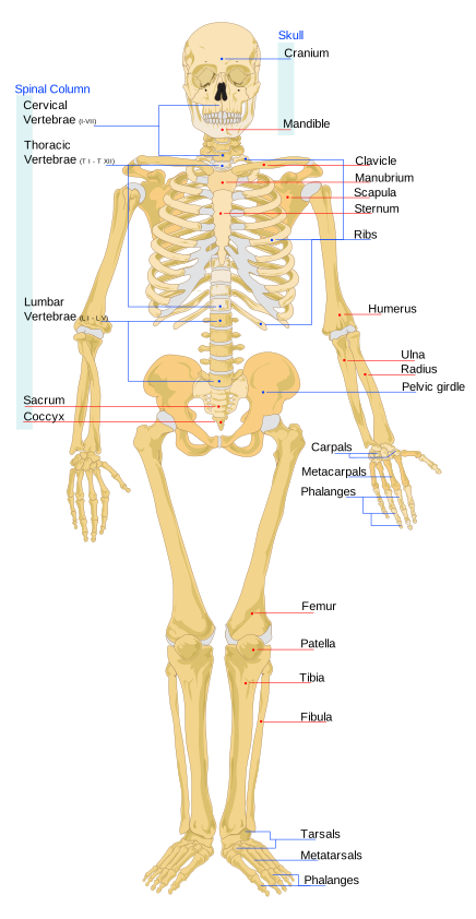
This topic covers bone shapes and bone cells, the two divisions of the skeleton, how joints are classified, the three muscle tissues, the steps of muscle contraction, and a few key muscles and their actions. Learn each structure together with its job, because most questions ask you to match the two.
Bone shapes and bone structure
- Long bones are longer than they are wide: femur, tibia, fibula, humerus, radius, ulna, metacarpals, metatarsals and phalanges.
- Short bones are cube-like: the carpals of the wrist and the tarsals of the ankle.
- Flat bones are thin: the sternum, ribs, scapulae and cranial bones.
- Irregular bones have complex shapes: the vertebrae and many facial bones.
- Sesamoid bones are small bones embedded in tendons, such as the patella (kneecap).
- A long bone has a shaft (diaphysis) around a medullary cavity of yellow marrow, and wider ends (epiphyses) filled with spongy bone.
- The epiphyseal (growth) plate is a layer of cartilage where a long bone lengthens; it becomes the epiphyseal line when growth stops.
- The periosteum is the fibrous membrane on the outer surface of a bone, where muscles and tendons attach.
Bone cells and bone tissue
- Osteogenic (osteoprogenitor) cells are the stem cells of bone; they develop into osteoblasts.
- Osteoblasts build new bone by secreting collagen matrix and calcium salts.
- Osteocytes are mature bone cells, former osteoblasts walled into small spaces (lacunae), that help maintain the surrounding matrix.
- Osteoclasts are large, multinucleated cells, derived from monocytes and macrophages, that break bone down (resorption).
- Compact bone is made of osteons (Haversian systems), each a set of concentric rings of mineralized matrix (lamellae) surrounding a central canal for blood vessels and nerves.
- Spongy bone is a lattice of struts called trabeculae, and its spaces hold red marrow, where blood cells form.
Axial and appendicular skeleton
- The adult skeleton has 206 bones: 80 axial and 126 appendicular.
- The axial skeleton is the skull, the hyoid and middle-ear bones, the vertebral column (vertebrae, sacrum and coccyx) and the thoracic cage (ribs and sternum).
- The appendicular skeleton consists of the limb bones and the girdles that connect the limbs to the axial skeleton.
- The pectoral girdle is the scapula and clavicle, so both are appendicular, not axial.
Joints
- By movement (function): a synarthrosis is immobile, an amphiarthrosis is slightly movable, and a diarthrosis is freely movable.
- By structure: fibrous joints (skull sutures), cartilaginous joints (pubic symphysis, intervertebral discs) and synovial joints (fluid-filled joint cavity).
- Skull sutures are fibrous synarthroses; the pubic symphysis and intervertebral discs are cartilaginous amphiarthroses; every synovial joint is a diarthrosis.
- A synovial joint has articular cartilage on the bone ends, an articular capsule, and a synovial membrane that secretes lubricating fluid.
- Ball-and-socket joints (shoulder and hip) move around many axes; the shoulder has the greater range of motion and the hip the greater stability.
- Hinge joints (elbow, knee, ankle, finger joints) bend and straighten along one axis.
- Pivot joints (between the first two vertebrae, and the proximal radioulnar joint) allow rotation.
- Condyloid joints (wrist and knuckles) and the saddle joint at the base of the thumb move in two planes.
- Plane joints, such as those between the carpal bones, allow small gliding movements.
The three muscle tissues
- Skeletal muscle: long cylindrical fibers, striated, many nuclei at the edge of each fiber, voluntary, attached to bones.
- Cardiac muscle: short branched cells, striated, usually one central nucleus, involuntary, found only in the heart.
- Smooth muscle: spindle-shaped cells, not striated, one nucleus, involuntary, found in the walls of organs and passageways such as blood vessels.
- Intercalated discs link cardiac cells end to end: desmosomes anchor neighboring cells, and gap junctions let electrical signals cross so the heart beats as one unit.
How a skeletal muscle contracts
The sarcomere, running from one Z disc to the next, is the contracting unit. Thick filaments are myosin; thin filaments are actin, with the regulatory proteins troponin and tropomyosin.
- A motor neuron releases acetylcholine at the neuromuscular junction, and an action potential spreads along the muscle fiber.
- The signal makes the sarcoplasmic reticulum let calcium ions out into the cell.
- Calcium binds troponin, which shifts tropomyosin off the myosin-binding sites on actin.
- Myosin heads bind actin to form cross-bridges, and release of phosphate triggers the power stroke that pulls actin toward the center (M-line).
- A new ATP binding to myosin makes the head let go of actin; splitting that ATP re-cocks the head for the next cycle.
- Without ATP, myosin cannot detach, which is why muscles stiffen in rigor mortis.
- To relax, the sarcoplasmic reticulum pumps the calcium back in, and tropomyosin covers the binding sites again.
- During contraction the Z discs move closer and the I band narrows, but the A band (the length of the thick filaments) stays the same width, because the filaments slide rather than shorten.
Muscles that work together
- The prime mover (agonist) produces a movement, the antagonist does the opposite action, and synergists assist the prime mover.
- The diaphragm is the prime mover of inhalation; the external intercostals assist by raising the ribs, and the internal intercostals pull the ribs together for forced exhalation.
- The rectus abdominis flexes the vertebral column, as in a sit-up.
- The quadriceps femoris on the front of the thigh extends the knee, as in kicking.
- The hamstrings on the back of the thigh flex the knee and extend the thigh at the hip.
- The gluteus maximus extends the thigh at the hip.
- The gastrocnemius and soleus plantar flex the foot (point the toes), and the tibialis anterior dorsiflexes it.
Key terms
- Long bone
- Any skeletal piece longer than it is wide, such as the femur or humerus.
- Osteoblast
- Bone-forming cell that secretes collagen matrix and calcium salts.
- Osteoclast
- Large multinucleated cell that breaks down bone matrix and releases its minerals.
- Osteocyte
- Mature bone cell in a lacuna that helps maintain the surrounding matrix.
- Osteon (Haversian system)
- Structural unit of compact bone: rings of lamellae around a central canal.
- Trabeculae
- Lattice of bony struts that forms spongy bone and holds red marrow.
- Appendicular skeleton
- The limb bones plus the pectoral and pelvic girdles that attach them to the trunk.
- Axial skeleton
- Skull, hyoid, vertebral column and thoracic cage, forming the body's central framework.
- Synarthrosis
- An immobile joint, such as a skull suture.
- Diarthrosis
- A freely movable joint; every synovial joint is one.
- Ball-and-socket joint
- A rounded bone end set in a cup-shaped cavity, allowing movement around many axes; the shoulder and hip.
- Intercalated disc
- Junction joining cardiac cells, with desmosomes for strength and gap junctions for electrical signals.
- Sarcomere
- Contracting unit of a muscle fiber, from one Z disc to the next.
- Troponin
- Thin-filament protein that binds calcium and moves tropomyosin off actin's binding sites.
- Cross-bridge
- Attachment of a myosin head to actin that pulls the thin filament during contraction.
- Prime mover
- The muscle mainly responsible for a given movement; also called the agonist.
- Diaphragm
- Dome-shaped muscle under the lungs; the prime mover of inhalation.
- Quadriceps femoris
- Four muscles on the front of the thigh that extend the knee.
Common mistakes
- Osteoblasts build bone and osteoclasts break it down; osteocytes maintain mature bone, and osteogenic cells are the stem cells that become osteoblasts.
- Trabeculae belong to spongy bone; the osteon is the unit of compact bone, and the periosteum and growth plate are not microscopic units at all.
- The scapula and clavicle are appendicular (pectoral girdle), while the sternum, sacrum and hyoid are axial.
- Synarthrosis, amphiarthrosis and diarthrosis are functional classes; symphysis, suture and synovial describe structure, so do not mix the two systems.
- Striations are not unique to the heart (skeletal muscle has them too), and many peripheral nuclei and voluntary control describe skeletal muscle; intercalated discs are the cardiac-only feature.
- Calcium binds troponin, not tropomyosin, myosin or actin; and ATP binding makes myosin detach, while the power stroke follows phosphate release.
Checked against: Wikipedia, "Bone"; Wikipedia, "Long bone"; Wikipedia, "Short bone"; Wikipedia, "Flat bone"; Wikipedia, "Irregular bone"; Wikipedia, "Sesamoid bone"; Wikipedia, "Epiphyseal plate"; Wikipedia, "Periosteum"; Wikipedia, "Bone marrow"; Wikipedia, "Osteoblast"; Wikipedia, "Osteocyte"; Wikipedia, "Osteoclast"; Wikipedia, "Osteon"; Wikipedia, "Haversian canal"; Wikipedia, "Trabecula"; Wikipedia, "Human skeleton"; Wikipedia, "Axial skeleton"; Wikipedia, "Appendicular skeleton"; Wikipedia, "Joint"; Wikipedia, "Cartilaginous joint"; Wikipedia, "Synovial joint"; Wikipedia, "Ball-and-socket joint"; Wikipedia, "Shoulder joint"; Wikipedia, "Hip"; Wikipedia, "Hinge joint"; Wikipedia, "Pivot joint"; Wikipedia, "Condyloid joint"; Wikipedia, "Saddle joint"; Wikipedia, "Muscle tissue"; Wikipedia, "Skeletal muscle"; Wikipedia, "Striated muscle tissue"; Wikipedia, "Smooth muscle"; Wikipedia, "Intercalated disc"; Wikipedia, "Muscle contraction"; Wikipedia, "Sarcomere"; Wikipedia, "Rigor mortis"; Wikipedia, "Anatomical terms of muscle"; Wikipedia, "Thoracic diaphragm"; Wikipedia, "Intercostal muscles"; Wikipedia, "Rectus abdominis muscle"; Wikipedia, "Quadriceps femoris muscle"; Wikipedia, "Hamstring"; Wikipedia, "Gluteus maximus"; Wikipedia, "Triceps surae muscle"; Wikipedia, "Tibialis anterior muscle"