This topic covers how the nervous system is organized, how neurons signal, what each brain region does, spinal reflexes, the sympathetic and parasympathetic divisions, the ear and eye, and the main endocrine glands and hormones. Most questions ask you to match a structure or hormone to its job, so learn them in pairs.
Organization of the nervous system
- The CNS (central nervous system) consists of the brain plus the spinal cord.
- Everything nervous outside them belongs to the PNS (peripheral nervous system): cranial nerves, the 31 pairs of spinal nerves, and ganglia.
- The somatic division serves skeletal muscle and conscious sensation; the autonomic division runs smooth muscle, cardiac muscle and glands largely without conscious control.
- Gray matter is rich in neuron cell bodies and dendrites; white matter is mainly myelinated axons.
- Inside the CNS a bundle of axons is called a tract; outside it, axon bundles make up nerves.
Neurons and glial cells
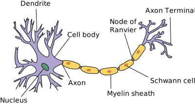
- Dendrites take in signals, the cell body contains the nucleus, and the axon conducts the impulse out to the axon terminals.
- Myelin is a lipid-rich insulating wrap on axons; the bare gaps between its segments are the nodes of Ranvier.
- Schwann cells form PNS myelin, and each one covers a stretch of just one axon.
- Oligodendrocytes form CNS myelin, and a single one can wrap stretches of several axons.
- Astrocytes control the chemical surroundings of CNS neurons and help build the blood-brain barrier.
- Microglia act as the CNS's immune cells, engulfing debris, dead cells and pathogens.
- Ependymal cells line the brain's ventricles and help produce cerebrospinal fluid.
Nerve impulses and synapses
- At rest the inside of a neuron sits near −70 mV, and the sodium-potassium pump spends ATP to maintain the ion gradients.
- When threshold is reached (about −55 mV), voltage-gated sodium channels open and Na⁺ floods in (depolarization).
- Next, voltage-gated potassium channels open and K⁺ moves out (repolarization).
- An action potential is all or none: a stronger stimulus raises how often impulses fire, not how large they are.
- Along myelinated axons the impulse leaps from node to node (saltatory conduction), which speeds it up.
- When the impulse arrives at the axon terminal, voltage-gated calcium channels open and Ca²⁺ flows in.
- Calcium makes the vesicles merge with the terminal membrane and empty neurotransmitter into the synaptic cleft (exocytosis).
- The neurotransmitter attaches to receptors on the next cell; some of these are ligand-gated ion channels that then open.
- The signal stops when the neurotransmitter is broken down (acetylcholinesterase splits acetylcholine), taken back up, or diffuses away.
The brain
- Frontal lobe: primary motor cortex (voluntary movement), speech production (Broca's area) and planning.
- Parietal lobe: sensing touch, pain and body position.
- Temporal lobe: hearing and memory; occipital lobe: processing vision.
- The thalamus passes sensory signals (all except smell) on to the cerebral cortex.
- The hypothalamus oversees homeostasis and steers the autonomic nervous system and the pituitary gland.
- The cerebellum uses input from muscles, joints and balance receptors to fine-tune movement, keeping it smooth, well timed and coordinated; it does not start movement.
- The medulla oblongata, working with the pons, controls heart rate, blood vessel diameter and breathing, so brain stem damage can be life-threatening.
Spinal cord and reflexes
- Sensory axons reach the spinal cord through the dorsal (posterior) root, and their cell bodies lie in the dorsal root ganglion.
- Motor axons exit the spinal cord through the ventral (anterior) root.
- In the gray matter, the posterior horn processes sensory input and the anterior horn contains motor neurons for skeletal muscle.
- A spinal reflex arc runs receptor → sensory neuron → spinal cord (often via an interneuron) → motor neuron → muscle, with no need to wait for the cerebrum.
- In the stretch reflex (such as the knee jerk), stretch detected by a muscle spindle makes that same muscle contract, while the opposing muscle's motor neurons are inhibited so it relaxes.
- In the withdrawal reflex, a painful stimulus makes the limb jerk away from it.
Autonomic nervous system
Most organs are supplied by both divisions, which balance each other.
- Sympathetic (fight or flight): heart rate rises, pupils dilate, airways widen, sweating increases, and blood flow shifts toward skeletal muscle.
- The sympathetic division also holds back digestion and prompts the adrenal medulla to release epinephrine and norepinephrine.
- Parasympathetic (rest and digest): heart rate slows, pupils constrict, and digestion speeds up, mostly through the vagus nerve.
The ear and eye
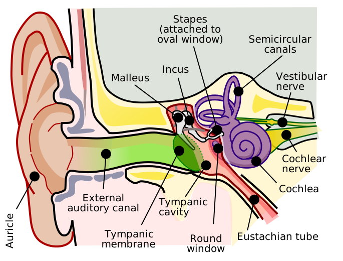
- Sound path: ear canal → tympanic membrane (eardrum) → ossicles (malleus, incus, stapes) → oval window → fluid of the cochlea.
- The organ of Corti, resting on the cochlea's basilar membrane, holds hair cells that turn these vibrations into nerve signals.
- The semicircular canals sense head rotation; the utricle and saccule sense head tilt and straight-line acceleration.
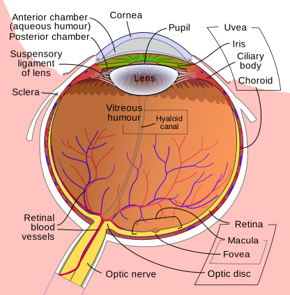
- The cornea and lens bend light so it focuses on the retina.
- Rods respond in dim light but give only grayscale vision; cones need brighter light and provide color vision.
- The fovea provides the sharpest vision, and the optic disc, where the optic nerve leaves, has no photoreceptors and forms a blind spot.
Endocrine glands and hormones
- The hypothalamus regulates the anterior pituitary through releasing and inhibiting hormones.
- The anterior pituitary secretes growth hormone, TSH (to the thyroid), ACTH (to the adrenal cortex), FSH and LH (to the gonads) and prolactin (milk production).
- The posterior pituitary makes no hormones of its own; it stores and releases ADH and oxytocin produced in the hypothalamus.
- ADH makes the kidneys reabsorb more water; oxytocin drives uterine contractions in labor and triggers milk letdown.
- The thyroid makes T3 and T4 (which require iodine and raise metabolic rate) and calcitonin (which lowers blood calcium).
- Parathyroid hormone raises blood calcium, partly by indirectly stimulating osteoclasts to break down bone.
- The adrenal cortex makes aldosterone (sodium retention) and cortisol (long-term stress, raises blood glucose); the adrenal medulla makes epinephrine and norepinephrine.
- Pancreatic beta cells release insulin, which lowers blood glucose by increasing cells' uptake of it; alpha cells release glucagon, which raises it.
- Glucagon, cortisol and epinephrine all raise blood glucose; of these four hormones, insulin is the only one that lowers it.
Key terms
- Central nervous system
- The brain and spinal cord.
- Peripheral nervous system
- All nerves and ganglia outside the brain and spinal cord.
- Schwann cell
- Peripheral glia that forms myelin around one segment of a single axon.
- Oligodendrocyte
- Glial cell that myelinates CNS axons, often several axons at once.
- Myelin
- Lipid-rich wrapping around axons that insulates them and makes impulses travel faster.
- Action potential
- All-or-none electrical impulse: Na⁺ moves in to depolarize, then K⁺ moves out to repolarize.
- Synapse
- Contact point where a neuron signals another cell, usually by releasing a neurotransmitter.
- Cerebellum
- Brain region that coordinates movement and balance, fine-tuning motor activity with sensory input.
- Medulla oblongata
- Lowest part of the brain stem, with centers that control heart rate, blood pressure and breathing.
- Hypothalamus
- Brain region that runs homeostasis and directs the autonomic nervous system and pituitary gland.
- Dorsal root
- Spinal nerve branch carrying sensory axons into the spinal cord.
- Ventral root
- Spinal nerve branch carrying motor axons out of the spinal cord.
- Reflex arc
- Receptor, sensory neuron, spinal cord connection, motor neuron and effector producing an automatic response.
- Sympathetic division
- Fight-or-flight branch: speeds the heart, dilates pupils and airways, slows digestion.
- Parasympathetic division
- Rest-and-digest branch: slows the heart, constricts pupils, stimulates digestion.
- Organ of Corti
- Structure on the cochlea's basilar membrane whose hair cells turn sound vibrations into nerve signals.
- Anterior pituitary
- Gland that secretes GH, TSH, ACTH, FSH, LH and prolactin under hypothalamic control.
- Posterior pituitary
- Stores and releases ADH and oxytocin, which the hypothalamus makes.
- Insulin
- Hormone from pancreatic beta cells that lowers blood glucose by increasing cells' glucose uptake.
- Glucagon
- Hormone from pancreatic alpha cells that raises blood glucose by making the liver release glucose.
Common mistakes
- The spinal cord, cerebellum and brain stem are all CNS; only nerves and ganglia, such as a spinal nerve, belong to the PNS.
- Schwann cells myelinate PNS axons and oligodendrocytes myelinate CNS axons; astrocytes and microglia make no myelin.
- Calcium entering the axon terminal triggers neurotransmitter release; sodium and potassium movements carry the impulse, and acetylcholinesterase acts only after release.
- The cerebellum coordinates movement and balance, the medulla controls heart rate and breathing, the thalamus relays sensation, and the hypothalamus runs homeostasis.
- Dorsal means sensory in and ventral means motor out; and in a stretch reflex the antagonist is inhibited and relaxes, handled in the spinal cord.
- TSH comes from the anterior pituitary, not the thyroid or hypothalamus; ADH is made in the hypothalamus and only released from the posterior pituitary; and of glucagon, cortisol, epinephrine and insulin, only insulin lowers blood glucose.
Checked against: Wikipedia, Nervous system; Wikipedia, Spinal nerve; Wikipedia, Nerve tract; Wikipedia, Neuron; Wikipedia, Myelin; Wikipedia, Neuroglia; Wikipedia, Schwann cell; Wikipedia, Action potential; Wikipedia, Sodium–potassium pump; Wikipedia, Chemical synapse; Wikipedia, Acetylcholinesterase; Wikipedia, Lobes of the brain; Wikipedia, Broca's area; Wikipedia, Temporal lobe; Wikipedia, Thalamus; Wikipedia, Hypothalamus; Wikipedia, Cerebellum; Wikipedia, Brainstem; Wikipedia, Medulla oblongata; Wikipedia, Spinal cord; Wikipedia, Posterior grey column; Wikipedia, Dorsal root ganglion; Wikipedia, Reflex arc; Wikipedia, Stretch reflex; Wikipedia, Withdrawal reflex; Wikipedia, Autonomic nervous system; Wikipedia, Ear; Wikipedia, Organ of Corti; Wikipedia, Vestibular system; Wikipedia, Human eye; Wikipedia, Blind spot (vision); Wikipedia, Pituitary gland; Wikipedia, Vasopressin; Wikipedia, Oxytocin; Wikipedia, Thyroid; Wikipedia, Parathyroid hormone; Wikipedia, Adrenal gland; Wikipedia, Cortisol; Wikipedia, Insulin; Wikipedia, Glucagon; Wikipedia, Blood sugar regulation